The symptoms of the leukoplakia of the buccal membranes and lips at its initial stage are analyzed on the basis of cytological data in parallel comparison with the data of fluorescent, histological and histochemical studies. The characteristic patterns of the individual forms of the disease in their first stage of development should be identified in order to formulate each form of recommendations for treatment to the patient.
In the process of the treatment, it is important to identify the conditions prior to the clinical course of each leukoplakia form. An objective examination allows the dentist to distinguish the specific changes in the state of the mucosa» – explain dentists in Indiana.
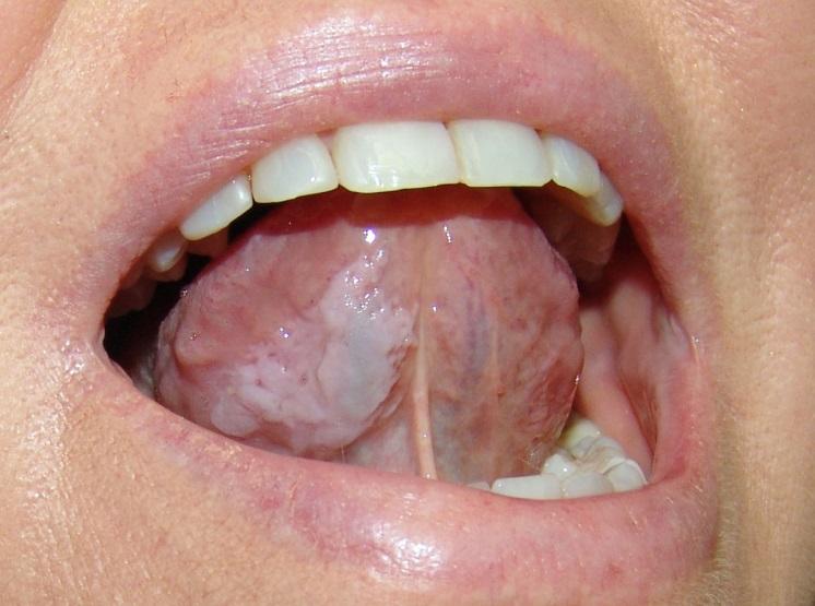
The loss of luster of the mucous membranes, prior keratosis is also pointed out by some clinicians. The dimness of the mucosa can be regarded as the first stage of leukoplakia.
The primary symptom of leukoplakia is the chronic inflammation and modified buccal mucosa epithelium. Clinical diagnosis of the initial form of leukoplakia includes the localization of the inflamed areas on the mucous membrane of the cheeks, lips and palate. If it affects the tongue, the clinical manifestations of this form are identified with difficulty and gets difficult to diagnose as well. And, finally, the diagnosis is possible only after the additional physical and laboratory methods which are carried out.
Subjective feelings at the initial stage of leukoplakia development are unchanged. Few patients notice a whitish color of individual areas on the oral mucosa (eg, on the lip mucosa).
When the shiny surface of the buccal mucosa on the line between the teeth, close to the corner of the mouth becomes dim, the affected area is found easily then. In this area, the oral mucosa has lost the glow, and its color has changed into greyish, with elements of keratinization present on the surface.
There is a slight increase in leukocyte emigration reaction of the mucous membranes of the whole mouth, without exception, in the patients with the primary form of leukoplakia.
Histological examinations show the moderate form of keratosis, para-and hyperkeratosis, or the intermittent type of keratinization. On the upper layer of mucous membrane nipples remain. Meanwhile, acanthosis is not pronounced. In hyperkeratosis, the granular layer cells are elongated and contain eleidin grains, which later develop into keratinized layer with flat cells.
The treatment objective at the initial stage is to eliminate the causal factors contributing to the development of the disease and to achieve the full mouth rehabilitation. The prescription of vitamins A, D, B is usually made to normalize the keratinization processes and is effective. Necessary hygiene measures are to be also taken during this disease, which becomes vital.
If you need qualitative treatment offered by certified dental experts, don’t forget to visit this page ttp://dentalprofy.com/dentists/alaska where you get treated from some of the renowned and well experienced dentists. It is always better to get your dental consultation done regularly so that you are well aware of your condition before time.